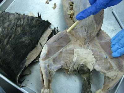
The probe is pointing to the pectineus muscle (anterior view) under the gracilis (cut and retracted).
Thursday, September 29, 2011
Cat Muscles - pectineus (3 of 3 muscles under gracilis)
The probe is pointing to the pectineus muscle (anterior view) under the gracilis (cut and retracted).
Cat Muscles - adductor longus (2 of 3 muscles under gracilis)

The probe is pointing to the adductor longus muscle (anterior view) under gracilis (cut and retracted).
Cat Muscles - adductor femoris (1 of 3 muscles under gracilis)
Wednesday, September 28, 2011
Cat Muscles - vastus medialis (2 of 3 thigh muscles under sartorius)
Cat Muscles - rectus femoris (1 of 3 thigh muscles under sartorius)
Cat Muscles - tibialis anterior
In this (approximately) anterior view, the probe is pointing to the tibialis anterior muscle, which is mostly located on the lateral border of the hindleg (below the thigh), though it is still technically on the anterior surface as well. Here is another picture of tibialis anterior on a cat from a *posterior* view (spine-up side).
Cat Muscles - gracilis
The probe is pointing to the gracilis muscle in the groin region on the cat (anterior view). Here's a silly, but possibly effective memory hook (works for me): "there's more 'grace'-ilis in being *next to* a cat's crotch" (than to actually be its crotch).
Cat Muscles - soleus (part of true "calf")
This probe (posterior view of the cat's hindleg) is pointing to the soleus muscle of what is technically part of the "calf muscle" (commonly thought of as only the gastrocnemius, but that is actually composed of 3 muscles, which includes the soleus). The soleus muscle is located immediately deep to the gastrocnemius. In this photo, you can clearly see that the most top/most superficial layer shown (gastrocnemius) inserts, and is even flush with, the calcaneal tendon ("Achilles heel"). Since this is an obvious the hallmark for the gastrocnemius, you can just count 1 layer deep to it and be sure it is the soleus muscle.
Cat Muscles - gastrocnemius ("calf muscle")
This is a posterior view of the cat's hindleg in which the probe is pointing to the gastrocnemius muscle or "stomach of the leg" (aka "calf muscle", although in anatomical terms the calf muscle is made up of 3 muscles: gastrocnemius, plantaris, and the soleus muscle). The gastrocnemius is the largest muscle on the "shank" (i.e. the area between the knee and the ankle on a human, which corresponds to the cat's thigh region). The gastrocnemius is immediately inferior to the hamstring group (biceps femoris needed to be only slightly retracted to reveal the upper part of gastrocnemius).
Cat Muscles - semitendinous (3 of 3 in "hamstring" muscle group)
In this anterior view, the probe is pointing to the semitendinosus
muscle of the hamstring group (again, made up of 3 muscles: biceps femoris, semimembranosus, and semitendinosus muscles). The biceps femoris
muscle (top layer) has been cut and retracted to reveal the
semitendinosus muscle that is deep to it. The semitendinosus muscle is located the closest to the cat's tail relative to the other 2 hamstring muscles, and is also inferior to semimembranosus muscle. Memory hook: "semi-T-endinosus is next to the T-ail of the cat".
Cat Muscles - semimembranosus (2 of 3 in "hamstring" muscle group)
In this anterior view, the probe is pointing to the semimebranosus
muscle of the hamstring group (made up of 3 muscles). The biceps femoris
muscle (top layer) has been cut and retracted to reveal the
semimebranosus muscle that is deep to it. The semimembranosus muscle is
in the "middle" of the 3-muscle hamstring group located on the posterior
surface of the hindleg.
Cat Muscles - biceps femoris (1 of 3 in "hamstring" muscle group)
Cat Muscles - fascia lata (*NOT a muscle, but a tendon/aponeurosis)
The probe is pointing to the fascia lata, which is the tendon/aponeurosis associated with the tensor fascia latae muscle of the thigh and is also known as the "deep fascia" of the thigh. The fascia lata aponeurosis is located posterior to the sartorius muscle, on the lateral
surface of the thigh.
To find fascia lata, first locate where the thigh joins the abdomen, then follow the connective tissue band that is on the lateral side of the thigh (from an anterior view): this is the fascia lata. In other words, the fascia lata aponeurosis of the cat's thigh is recognizable as the tough and semi-transparent sheet of connective tissue (fascia) that extends downward from a triangular muscle, the tensor fascia latae.
To find fascia lata, first locate where the thigh joins the abdomen, then follow the connective tissue band that is on the lateral side of the thigh (from an anterior view): this is the fascia lata. In other words, the fascia lata aponeurosis of the cat's thigh is recognizable as the tough and semi-transparent sheet of connective tissue (fascia) that extends downward from a triangular muscle, the tensor fascia latae.
Cat Muscles - tensor fascia latae (sheet-like muscle)
The probe is pointing to the tensor fascia latae muscle, which is a triangular muscle* that can be found at the junction of the abdomen with the thigh (anterior view). The tensor fascia latae muscle narrows as it approaches its insertion into the fascia lata aponeurosis (a sheet-like tendon).
*you can only see a VERY small portion of this muscle in the photo above.
Thursday, September 22, 2011
Cat Muscles - brachioradialis
 The probe is lodged THROUGH the brachioradialis. IMPORTANT: The photo view is from the cat being positioned on its belly ("prone" position).
Description of brachioradialis - is a thin, string-like muscle on the lateral side of the humerus (on the side that is farther away from the ribcage).
Function of brachioradialis - flexes the elbow joint.
Cat Muscles - sartorius (aka "the tailor's muscle")
The probe is pointing to the sartorius muscle on the leg of the cat (hindlimb).
Description of sartorius - is the most anterior (belly-up side) muscle of the cat thigh. The sartorius looks like a wide, thin band extending from its origin on the ileum (of the pelvis) to its insertion point on the patella (knee) and tibia. The majority of the muscle covers the medial side of the thigh, covering nearly half of the anterior (again, belly-side) surface. This can be remembered as the "tailor's muscle" (sartor = "tailor) because it is traditionally the muscle area where a tailor measures a client's inseam for fitting pants.
Function of sartorius - adducts and rotates the thigh bone.
Cat Muscles - biceps brachii
 The probe is pointing to biceps brachii.
Description of biceps brachii - is a large spindle-shaped muscle (i.e. tapers at both ends) that can be seen when the epitrochlearis flap (cut) is retracted (pulled back). The biceps brachii muscle's location is superior to the triceps brachii muscle on the humerus of the cat. This muscle is more prominent in humans, but its origin, insertion point, and function/action are very similar in humans.
Function of biceps brachii - flexes forearm.
Cat Muscles - triceps brachii
 The probe is pointing to triceps brachii.
Description of triceps brachii - is the large, fleshy muscle on the upper arm (humerus) that is located on the surface of the humerus closer to the side of the body. This muscle can be seen when the epitrochlearis flap (cut) is retracted (pulled back).
Function of triceps brachii - extends the forearm (radius and ulna).
Cat Muscles - epitrochlearis (there is NO human equivalent)
 The probe is pointing to a flap we made by cutting a piece of the upper arm/proximal end of the humerus.
Description of epitrochlearis - is a broad, flat, and extremely THIN muscle that has its origin (a tendon) on the latissimus dorsi and inserts into the ulna.
Function of the epitrochlearis - extends the forelimb (radius & ulna) of the cat.
Cat Muscles - serratus ventralis
 A different cat specimen (definitely not our cat, Donkey J. Meowmers) was used as a representative for serratus ventralis because it had a clearer "pocket" for this muscle. Although the photo here does not show the "serrated" or fingerlike pieces of muscle (3-4 "sections") in the dark space indicated by the gloved finger, this is the location of the serratus ventralis. To fully expose this muscle, we would have to cut open* the latissimus dorsi (upper rib/armpit "wings").
Description of serratus ventralis - in the cat, the serratus ventralis is one muscle, while the analogous muscle in humans is 2 separate muscles (serratus anterior and serratus posterior).
*Updated picture to come later, possibly!
Cat Muscles - internal oblique
 The probe in the picture (above) is pointing to the internal oblique sheet of muscle in the abdomen.
In both pictures (above), the probe points to the internal oblique sheet of muscle.
Description of internal oblique - located deep to the external oblique sheet of muscle, and also located beside the rectus abdominis (just like the external oblique).
Cat Muscles - external oblique

The probe is in the general area of the external oblique muscle fibers (cottony-white stuff).
Description of external oblique - is a sheet of muscle located directly on either side of to the rectus abdominis band of muscle. The external oblique sheet of muscle is superficial to and directly covers the internal oblique.
Wednesday, September 21, 2011
Cat Muscles - clavodeltoid (aka "deltoid" in humans)
 Probe is pointing to clavodeltoid muscle (above AND below)
Both pictures show an anterior view of the cat's clavodeltoid muscle (aka clavobrachialis), which is analogous to the human "deltoid" muscle. Simply think of this one as the "shoulder muscle" for the lab practical.
Description of clavodeltoid - it is the most superficial muscle of the shoulder. It's origin is the clavicle and its insertion point is the proximal end of the ulna of the forearm.
Function of clavodeltoid - draws the forearm toward the chest.
Cat Muscles - pectoantebrachialis (there is NO equivalent in humans)

The probe is pointing to the pectoantebrachialis muscle.
Description of pectoantebrachialis - a thin, straplike muscle that is superior to (vertically above) the pectoralis major muscle. This muscle is NOT found in humans, and there is not analogous muscle to it in humans either.
Function of pectoantebrachialis - just like the xiphihumeralis, pectoralis major, and pectoralis minor, this muscle helps draw the arm to the chest.
Cat Muscles - rectus abdominis (aka "6 pack")

The probe is pointing to rectus abdominis, which is a long and fairly wide BAND of muscle that runs along the midline of the body on the abdominal surface of the cat (or human). In this specimen, the rectus abdominis muscle is very white and prominent.
Fun fact: rectus abdominis is the muscle that very physically fit people/body builders develop into a "6 pack".
Cat Muscles* - linea alba (*NOT a muscle, just Connective Tissue)
 The probe is pointing to linea alba (aka "white line"), which is the very obvious vertical white "scar" that runs across the cat's body medially along the rectus abdominis.
Description/Function of linea alba - separates the rectus abdominis muscle into 2 (approximately) equal parts.
Cat Muscles - pectoralis major

The probe is pointing to the pectoralis major muscle.
Description of pectoralis major - the muscle is superior to (vertically above) pectoralis minor.
Function of pectoralis major - similar to the xiphihumeralis, the pectoralis major muscle inserts somewhere within the proximal half of the humerus AND helps bend the arm toward the chest.
Cat Muscles - xiphihumeralis

The probe is pointing to the xiphihumeralis muscle. It is directly inferior (vertically below) the pectoralis minor. In fact, it is closely associated with pectoralis minor in another way: xiphihumeralis's fibers run parallel to and are FUSED with the fibers of the pectoralis minor muscle.
Description of xiphihumeralis - this muscle's name tells you a few very important things about it, which makes things very convenient for us students! That is,
(1) xiphi- = refers to "xiphoid process" of the chest= origin of muscle
(2) -humeralis = refers to "humerus" (upper arm) = insertion point of muscle (proximal end of humerus)
The insertion point gives you the biggest clue to what the xiphihumeralis does...
Function of xiphihumeralis - helps draw the arm to the chest (pectoralis major and pectoralis minor also do this.)
Cat Muscles - pectoralis minor

Probe is pointing to pectoralis minor (yes, it is--even though it TOTALLY looks bigger than the other muscles that are sectioned off on the cat's chest, by scalpel cuts.
In this instance ONLY: Think of the "minor" name as the muscle being physically deeper down in the body than its pectoralis major counterpart. In fact, pectoralis minor is a muscle directly UNDERNEATH the pectoralis major muscle in 2 ways (in 2 planes): (1) it's inferior/vertically below AND (2) deep to the pectoralis major.
Don't confuse yourself by believing that "minor" and "major" refers to relative size--even though it DOES 99% of the time in anatomy (in Latin, minor = "small"; major = "large"). The Lab manual understands this dilemma, saying: "contrary to what its name implies, the pectoralis minor is a larger and thicker muscle than the pectoralis major".
Description of pectoralis minor - its origin is on the sternum and its insertion point is somewhere within the proximal half of the humerus.
Function of pectoralis minor - this muscle helps draw the arm to the chest (xiphihumeralis and pectoralis major also do this).
Cat Muscles - latissimus dorsi (anterior view only)

Note: gloved finger is NOT pointing to latissimus dorsi! The weird mangled "wing" of muscle directly to the RIGHT of the gloved finger is the latissimus dorsi.

Both pics (above) show anterior views of the latissimus dorsi muscle, which is basically the "wing"-like flap of muscle on top of where the upper ribs are on this cat (on the right side of each picture).
Description of latissimus dorsi: "The large flat muscle covering most of the lateral (side) surfaces of the posterior trunk." These pictures here won't help us visualize that--this is the anterior view only. However, you can see that if latissimus dorsi takes up this much space in FRONT of the body, then it must cover a whole lot of the back of it! It's similar in cats as in humans (latissimus dorsi is HUGE!), otherwise we would not be using cats to better understand human anatomy.
Cat Muscles - calcaneal tendon ("Achilles tendon")


 All 3 pictures (above) show the calcaneal tendon in the cat's foot (aka "Achilles tendon")--it's the super-taught band of tissue in the center of each picture. This is where the largest muscle on the "shank" of the cat's leg (aka "gastrocnemius") inserts into the calcaneus (in humans, known as the "heel bone" or tarsus bone).
 Description & Function of calcaneus tendon - The calcaneus tendon is made of strong, cord-like Connective Tissue (Dense Regular CT proper) that indirectly binds muscles to bones.
Cat Muscles - cutaneous maximus
 Pic 1 (above) : cutaneous maximus
Pic 2 (above): cutaneous maximus (Detail)
Description: Pic 1 and Pic 2 both show the "cutaneous maximus" muscle (the upper flap) on the cat. The Marieb Lab manual says that the cutaneous maximus is made up of a "thin layer of muscle fibers" that adhere to the (underside) of the skin of the cat.
Detail: In Pic 2, especially, you can clearly see "small, white cordlike structures extending from the flap of skin to the muscles at fairly regular intervals".
Function of cutaneous maximus: The cutaneous maximus "enables the cat to move its skin, rather like our facial muscles allow us to express emotion."
Subscribe to:
Comments (Atom)


